Figure 1
Rhabdomyoblasts in Pediatric Tumors: A Review with Emphasis on their Diagnostic Utility
Giuseppe Angelico*, Eliana Piombino, Giuseppe Broggi, Fabio Motta and Saveria Spadola
Published: 09 March, 2017 | Volume 1 - Issue 1 | Pages: 008-016

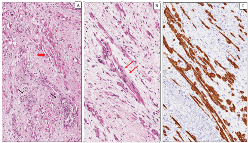
Figure 1:
Nephroblastoma (Wilms tumor). (A) Histological examination showing a proliferation of primitive tubular ad glomerular-like structures (black arrows) admixed with rabdomyoblats of variable size and shape (red arrow). (B) Elongated and oval rhabdomyoblasts with eccentric round nuclei and densely eosinophilic cytoplasm with occasional cytoplasmic cross-striations (arrows). (C) Diffuse and intense cytoplasmic immunoreactivity for desmin in the rhabdomyoblastic component.
Read Full Article HTML DOI: 10.29328/journal.jsctt.1001002 Cite this Article Read Full Article PDF
More Images
Similar Articles
-
Rhabdomyoblasts in Pediatric Tumors: A Review with Emphasis on their Diagnostic UtilityGiuseppe Angelico*,Eliana Piombino,Giuseppe Broggi,Fabio Motta,Saveria Spadola. Rhabdomyoblasts in Pediatric Tumors: A Review with Emphasis on their Diagnostic Utility . . 2017 doi: 10.29328/journal.jsctt.1001002; 1: 008-016
Recently Viewed
-
A Critical Review on Some Recent Developments in Comparison of Biological SequencesDK Bhattacharya*. A Critical Review on Some Recent Developments in Comparison of Biological Sequences. J Genet Med Gene Ther. 2024: doi: ; 7: 008-014
-
Advances in deep learning-based cancer outcome prediction using multi-omics dataAndrew Zhou, Charlie Zhang, Okyaz Eminaga*. Advances in deep learning-based cancer outcome prediction using multi-omics data. Ann Proteom Bioinform. 2023: doi: 10.29328/journal.apb.1001020; 7: 010-013
-
Phenotypic differences in Obese Patients with Heart Failure with Preserved Ejection Fraction (HFpEF) - A Mini ReviewMichelle Nanni*, Vivian Hu, Swagata Patnaik, Alejandro Folch Sandoval, Johanna Contreras. Phenotypic differences in Obese Patients with Heart Failure with Preserved Ejection Fraction (HFpEF) - A Mini Review. New Insights Obes Gene Beyond. 2024: doi: 10.29328/journal.niogb.1001020; 8: 001-005
-
The Stability and Behaviour of the Superposition of Non-Linear Waves in SpaceChristopher O Adeogun*. The Stability and Behaviour of the Superposition of Non-Linear Waves in Space. Int J Phys Res Appl. 2023: doi: 10.29328/journal.ijpra.1001075; 6: 216-221
-
Toxicity and Phytochemical Analysis of Five Medicinal PlantsJohnson-Ajinwo Okiemute Rosa*, Nyodee, Dummene Godwin. Toxicity and Phytochemical Analysis of Five Medicinal Plants. Arch Pharm Pharma Sci. 2024: doi: 10.29328/journal.apps.1001054; 8: 029-040
Most Viewed
-
Evaluation of Biostimulants Based on Recovered Protein Hydrolysates from Animal By-products as Plant Growth EnhancersH Pérez-Aguilar*, M Lacruz-Asaro, F Arán-Ais. Evaluation of Biostimulants Based on Recovered Protein Hydrolysates from Animal By-products as Plant Growth Enhancers. J Plant Sci Phytopathol. 2023 doi: 10.29328/journal.jpsp.1001104; 7: 042-047
-
Feasibility study of magnetic sensing for detecting single-neuron action potentialsDenis Tonini,Kai Wu,Renata Saha,Jian-Ping Wang*. Feasibility study of magnetic sensing for detecting single-neuron action potentials. Ann Biomed Sci Eng. 2022 doi: 10.29328/journal.abse.1001018; 6: 019-029
-
Physical activity can change the physiological and psychological circumstances during COVID-19 pandemic: A narrative reviewKhashayar Maroufi*. Physical activity can change the physiological and psychological circumstances during COVID-19 pandemic: A narrative review. J Sports Med Ther. 2021 doi: 10.29328/journal.jsmt.1001051; 6: 001-007
-
Pediatric Dysgerminoma: Unveiling a Rare Ovarian TumorFaten Limaiem*, Khalil Saffar, Ahmed Halouani. Pediatric Dysgerminoma: Unveiling a Rare Ovarian Tumor. Arch Case Rep. 2024 doi: 10.29328/journal.acr.1001087; 8: 010-013
-
Prospective Coronavirus Liver Effects: Available KnowledgeAvishek Mandal*. Prospective Coronavirus Liver Effects: Available Knowledge. Ann Clin Gastroenterol Hepatol. 2023 doi: 10.29328/journal.acgh.1001039; 7: 001-010

HSPI: We're glad you're here. Please click "create a new Query" if you are a new visitor to our website and need further information from us.
If you are already a member of our network and need to keep track of any developments regarding a question you have already submitted, click "take me to my Query."


