Figure 2
Rhabdomyoblasts in Pediatric Tumors: A Review with Emphasis on their Diagnostic Utility
Giuseppe Angelico*, Eliana Piombino, Giuseppe Broggi, Fabio Motta and Saveria Spadola
Published: 09 March, 2017 | Volume 1 - Issue 1 | Pages: 008-016
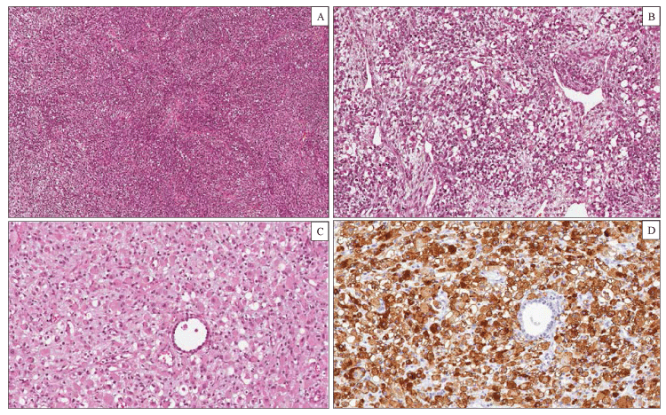
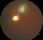
Figure 2:
Pleuropulmonary Blastoma type III. Histological examination showing a solid tumor composed of a mixture of blastemal and mesenchymal neoplastic cells (hematoxylin-eosin). (A) Large areas of the tumor consisted of spindle cells arranged in intersecting fascicles with a brosarcoma-like growth pattern. (B) Other elds of the tumor showed a proliferation of blastemal cells and rhabdomyoblasts in a myxoid stroma. (C) Rhabdomyoblasts displayed an oval shape, eccentric nuclei and a densely eosinophilic cytoplasm. (D) Diuse cytoplasmic immunohistochemical staining for desmin in rhabdomyoblastic foci.
Read Full Article HTML DOI: 10.29328/journal.jsctt.1001002 Cite this Article Read Full Article PDF
More Images
Similar Articles
-
Rhabdomyoblasts in Pediatric Tumors: A Review with Emphasis on their Diagnostic UtilityGiuseppe Angelico*,Eliana Piombino,Giuseppe Broggi,Fabio Motta,Saveria Spadola. Rhabdomyoblasts in Pediatric Tumors: A Review with Emphasis on their Diagnostic Utility . . 2017 doi: 10.29328/journal.jsctt.1001002; 1: 008-016
Recently Viewed
-
The Stability and Behaviour of the Superposition of Non-Linear Waves in SpaceChristopher O Adeogun*. The Stability and Behaviour of the Superposition of Non-Linear Waves in Space. Int J Phys Res Appl. 2023: doi: 10.29328/journal.ijpra.1001075; 6: 216-221
-
Toxicity and Phytochemical Analysis of Five Medicinal PlantsJohnson-Ajinwo Okiemute Rosa*, Nyodee, Dummene Godwin. Toxicity and Phytochemical Analysis of Five Medicinal Plants. Arch Pharm Pharma Sci. 2024: doi: 10.29328/journal.apps.1001054; 8: 029-040
-
Augmented and Virtual Reality in Forensic Odontology: Practical ImplementationsPiyush Asnani*, Shireen Ali. Augmented and Virtual Reality in Forensic Odontology: Practical Implementations. J Forensic Sci Res. 2023: doi: 10.29328/journal.jfsr.1001050; 7: 055-057
-
New Fungi Associated with Blackberry Root Rot (Rubus spp.), in Michoacán, MexicoLuis Mario Tapias Vargas, Anselmo Hernández Pérez, Adelaida Stephany Hernández Valencia*. New Fungi Associated with Blackberry Root Rot (Rubus spp.), in Michoacán, Mexico. J Plant Sci Phytopathol. 2024: doi: 10.29328/journal.jpsp.1001129; 8: 038-040
-
Sexual Dimorphism in Autoimmune DisordersPD Gupta*. Sexual Dimorphism in Autoimmune Disorders. Clin J Obstet Gynecol. 2024: doi: 10.29328/journal.cjog.1001164; 7: 056-058
Most Viewed
-
Evaluation of Biostimulants Based on Recovered Protein Hydrolysates from Animal By-products as Plant Growth EnhancersH Pérez-Aguilar*, M Lacruz-Asaro, F Arán-Ais. Evaluation of Biostimulants Based on Recovered Protein Hydrolysates from Animal By-products as Plant Growth Enhancers. J Plant Sci Phytopathol. 2023 doi: 10.29328/journal.jpsp.1001104; 7: 042-047
-
Feasibility study of magnetic sensing for detecting single-neuron action potentialsDenis Tonini,Kai Wu,Renata Saha,Jian-Ping Wang*. Feasibility study of magnetic sensing for detecting single-neuron action potentials. Ann Biomed Sci Eng. 2022 doi: 10.29328/journal.abse.1001018; 6: 019-029
-
Physical activity can change the physiological and psychological circumstances during COVID-19 pandemic: A narrative reviewKhashayar Maroufi*. Physical activity can change the physiological and psychological circumstances during COVID-19 pandemic: A narrative review. J Sports Med Ther. 2021 doi: 10.29328/journal.jsmt.1001051; 6: 001-007
-
Pediatric Dysgerminoma: Unveiling a Rare Ovarian TumorFaten Limaiem*, Khalil Saffar, Ahmed Halouani. Pediatric Dysgerminoma: Unveiling a Rare Ovarian Tumor. Arch Case Rep. 2024 doi: 10.29328/journal.acr.1001087; 8: 010-013
-
Prospective Coronavirus Liver Effects: Available KnowledgeAvishek Mandal*. Prospective Coronavirus Liver Effects: Available Knowledge. Ann Clin Gastroenterol Hepatol. 2023 doi: 10.29328/journal.acgh.1001039; 7: 001-010

HSPI: We're glad you're here. Please click "create a new Query" if you are a new visitor to our website and need further information from us.
If you are already a member of our network and need to keep track of any developments regarding a question you have already submitted, click "take me to my Query."


